

Eye Surgery Explained: What Happens During LASIK and Cataract Surgery
Cataract Surgery
Medical Perspective: During phacoemulsification, the surgeon creates a 2-3mm self-sealing clear corneal incision and performs a continuous curvilinear capsulorhexis to access the lens. Ultrasonic energy emulsifies the clouded crystalline lens, which is aspirated from the capsular bag. An intraocular lens is then implanted within the preserved posterior capsule, maintaining the natural barrier between anterior and posterior segments.
Everyday Perspective: Your eye's natural lens is about the size of a regular M&M candy with a thin outer shell. The surgeon makes a tiny opening (small enough to heal without stitches), creates a circular window in that shell, uses ultrasound energy to break up the cloudy lens inside, removes it, then slides a crystal-clear artificial lens into the same shell. We keep that protective coating intact to hold your new lens securely in place.
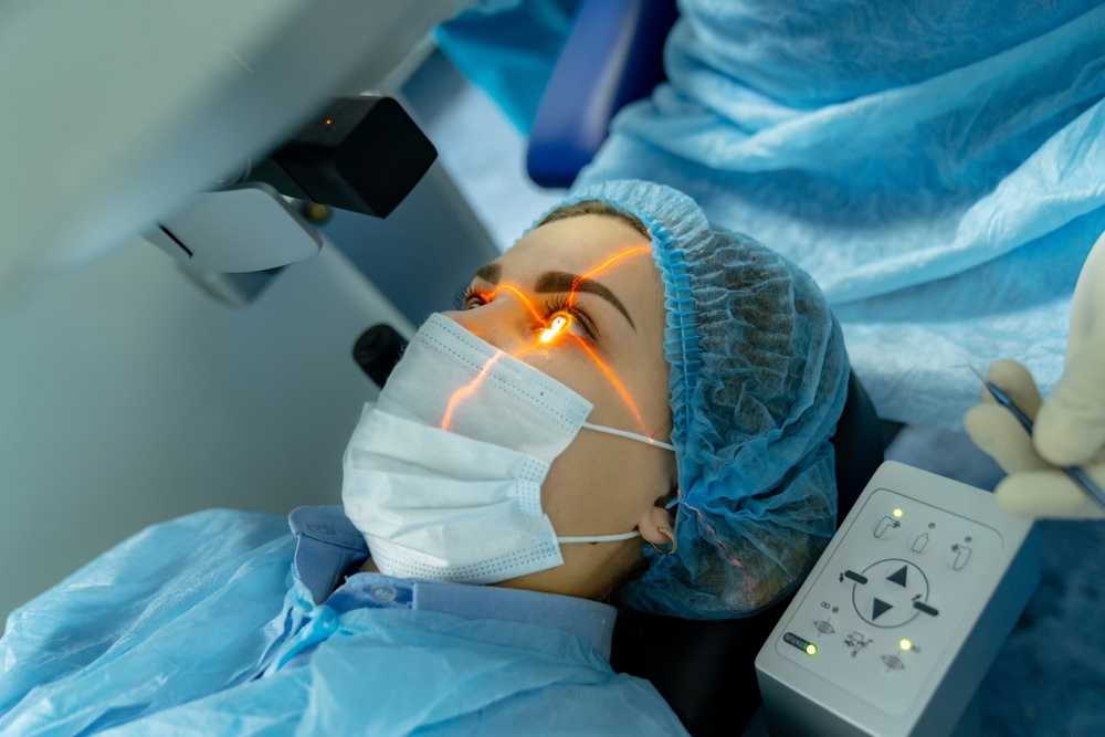
LASIK
Medical Perspective: LASIK involves creating a corneal flap and using excimer laser photoablation to reshape the stromal bed, correcting refractive error. Pre-operative pachymetry is critical - residual stromal bed thickness must remain ≥250-300 microns to prevent post-operative ectasia. Patients with insufficient corneal thickness are contraindicated due to biomechanical instability risk (such as corneal ectasia).
Everyday Perspective: Your cornea has layers like an Oreo - some people have thick "double-stuffed" corneas, others are thinner. Surgeons lift the top cookie layer, use a laser to remove precise amounts of the "cream filling" to correct your vision, then lay the cookie back down. The critical safety question: how much cream can we remove? Taking someone from double-stuffed down to a thin Oreo leaves the structure too weak, risking progressive bulging (ectasia). That's why thickness measurements determine if you're a safe candidate.
Quick Links

All Eye
Care Services

Keep
In Touch
MAP
© 2026 East Avenue Vision Center. All Rights Reserved. Accessibility Statement - Privacy Policy - Sitemap
Powered by: 